Olabisi O Ogunrinola ![]() ,
Dorcas A Wusu,
Olusegun O Fajana,
Samuel N Olaitan,
Zainab O Smith,
Abdul-Rahman I Bolaji
,
Dorcas A Wusu,
Olusegun O Fajana,
Samuel N Olaitan,
Zainab O Smith,
Abdul-Rahman I Bolaji
For correspondence:- Olabisi Ogunrinola Email: olabisi.ogunrinola@lasu.edu.ng Tel:+234803324476
Received: 16 March 2015 Accepted: 13 November 2015 Published: 29 January 2016
Citation: Ogunrinola OO, Wusu DA, Fajana OO, Olaitan SN, Smith ZO, Bolaji AI. Effect of low level cadmium exposure on superoxide dismutase activity in rat. Trop J Pharm Res 2016; 15(1):115-119 doi: 10.4314/tjpr.v15i1.16
© 2016 The authors.
This is an Open Access article that uses a funding model which does not charge readers or their institutions for access and distributed under the terms of the Creative Commons Attribution License (http://creativecommons.org/licenses/by/4.0) and the Budapest Open Access Initiative (http://www.budapestopenaccessinitiative.org/read), which permit unrestricted use, distribution, and reproduction in any medium, provided the original work is properly credited..
Purpose:To investigate the effect of low level cadmium (Cd) exposure on the activity of superoxide dismutase (SOD) in rat.
Methods:Thirty-two male albino rats were divided into four groups of eight animals each. Group one received distilled water and served as control. The other three groups were exposed to 100, 200 and 300 ppm cadmium doses, respectively, in their drinking water for 6 weeks. At the end of 6 weeks, blood, brain and liver were removed from the animals and SOD activities were investigated spectrophotometrically at 480 nm.
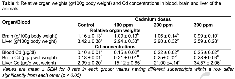
Results:The data revealed a significant (p < 0.05) decrease in organ weight of the exposed rats, and with the highest cadmium accumulation in liver ranging from 2.99 ± 0.20 µg Cd/g tissue in control to 34.57 ± 2.06 µg Cd/g tissue for the 300 ppm dose group. Exposure to cadmium resulted in significant (p < 0.05) decrease in SOD activity in plasma, erythrocytes, brain and liver in a dose-dependent manner compared to control. The data showed a negative correlation between tissue cadmium and SOD in plasma, erythrocyte and brain. While positive associations were observed between blood and tissue (brain and liver) cadmium and SOD, a significant (p < 0.01), negative correlation was found between tissue cadmium and SOD.
Conclusion:As Cd dose increase, plasma, erythrocyte, brain and liver SOD activities decrease. This might affect the maintenance of the normal body physiological conditions.
Introduction
Cadmium (Cd) is a serious environmental and occupational contaminant that may represent a serious health hazard to humans and other animals. The increased discharge of cadmium from industrial process, waste disposal and cigarette smoke in the environment leads to a general fear for the potential toxic effects of cadmium. Since cadmium cannot be degraded, the risk of environmental exposure is constantly increasing because of accumulation via the food chain [1]. Exposure to this toxic metal at the cellular level can produce both acute and chronic tissue injury and can damage various organs including lung, liver, kidney, bone, testis and placentas depending on the dosage, route and duration of exposure causing several damages [2,3]. The generation of reactive oxygen species (ROS) followed by development of oxidative stress in the target organs is one of the several mechanisms through which cadmium exerts its toxicity [4]. Vulnerability to stress alters the normal body homeostasis and leads to the development of various pathologies, which may involve alterations in the antioxidant defense system such as superoxide dismutase. Antioxidants may be enzymatic or non-enzymatic and are intimately involved in the prevention of cellular damage – the common pathway for cancer, aging and a diversity of diseases [5].
Superoxide dismutase is an important enzyme family in living cells for maintaining normal physiological conditions and for coping with stress. The role of superoxide dismutase, therefore, is to protect the biological integrity of the cells and tissues against harmful effects of superoxide free radical [6]. Since the implication of cadmium toxicity on disruption of the oxidative antioxidant defense system depends on route of administration, dose and duration of exposure, we propose to investigate the effect of low level of cadmium exposure through drinking water for six weeks on superoxide dismutase activity in adult male rat.
Methods
Animals and experimental design
Thirty-two adult male albino rats (Wistar strain) weighing between 180 to 200 g were purchased from Nigerian Institute of Medical Research, Yaba, Lagos, Nigeria. They were housed in animal stainless steel cages with a 12 h light/dark cycle and free access to food and water for 14 days prior to the experiment.
The animals were randomly and equally distributed into four groups with eight rats in each group. Animals in group one were given distilled water and served as controls while rats in three other groups were given 100, 200 and 300 ppm cadmium in drinking water, respectively. All groups were fed ad libitum with growers mash (product of Animal Care, Lagos, Nigeria). They were all exposed to this treatment for six weeks. At the end of the treatment period, the rats were weighed and sacrificed under light ether inhalation anesthesia. The blood was collected, centrifuge into plasma and erythrocyte while the brain and liver were quickly excised, weighed and placed on ice until required for biochemical analysis. All the animal protocols conformed to the guiding principles of laboratory animal care (NIH Publication No. 85-93, revised 1985) [7] and were approved by the adhoc animal ethical committee (ref no. BCH-05/2007) of the Department of Biochemistry, Lagos State University, Ojo, Lagos, Nigeria.
Superoxide dismutase assay
An indirect method of inhibiting auto- oxidation of epinephrine to its adrenochrome was used to assay superoxide dismutase (SOD) activity [8]. The 10 % homogenate, each of brain and liver, respectively, was prepared with chilled 0.9 % NaCl solution and centrifuged at 3,000 g for 10 min. The plasma, erythrocyte, homogenates of the brain and liver SOD was determined spectrophotometrically at 480 nm (model SM 755s, Surgienfield Instrument, Zhejiang, China. SOD activity was expressed in unit/ml for plasma and erythrocyte while that of the brain and liver homogenates were expressed in unit/g tissue. Erythrocyte SOD activity was assayed on the basis of the inhibition of adrenaline autoxidation. Briefly, to erythrocyte hemolysate, 333 μM solution of adrenalin in 50 mM carbonate, pH 10.2, containing 100 μM EDTA was added. The absorbance of the solution was monitored spectrophotometrically at 480 nm as a function of time at 37 ºC. The degree of inhibition of the maximal rate of absorbance increase was taken as a measure of SOD activity. One unit of SOD is the amount of the enzyme required for 50 % inhibition of the auto-oxidation of epinephrine to adenochrome per minute. The blood and organ cadmium concentrations were determined by atomic absorption spectrophotometry (Model NC 942340030042, product of Thermo Fisher Scientific Company, USA) after digestion with concentrated trioxonitrate (V) acid.
Statistical analysis
The assay was performed in triplicate and results were expressed as mean ± SEM. Statistical differences in the means were determined using one-way analysis of variance (ANOVA) followed by Turkey’s test (Turkey honest significant difference (THSD)) with p < 0.05 considered significant. The associations among the parameters and their magnitudes were tested for using multiple linear regression analysis.
Results
A gradual decrease was observed in the relative weights of the organs, although this was not dose dependent. At the highest dose of cadmium, relative weights of the organs decreased as follows: brain 15 % and liver 24 %, respectively. The result shows that cadmium uptake is high in blood, brain and liver of the animals, but the highest concentration was observed in the liver, whereas brain accumulated the least ().
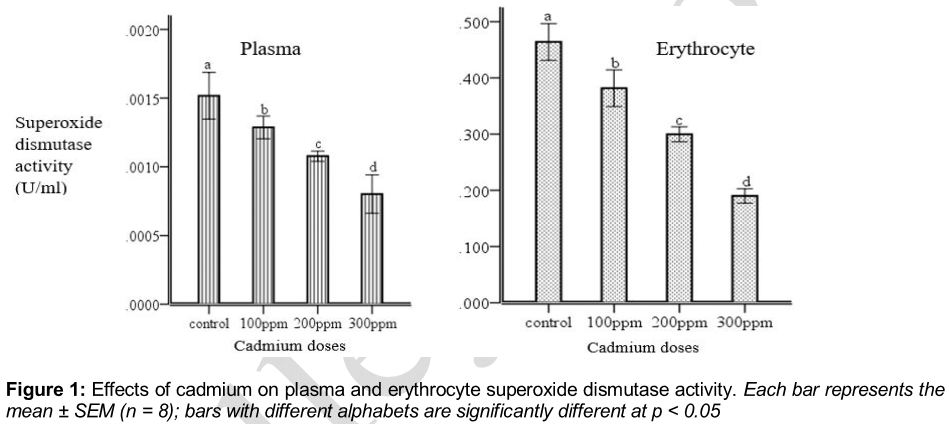
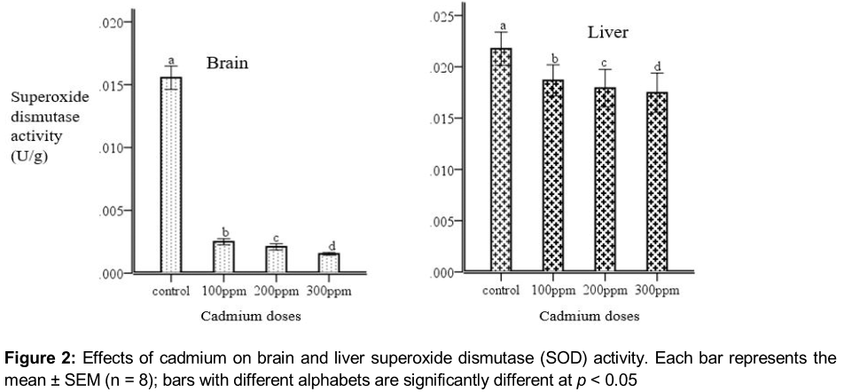
There was a dose-dependent reduction in plasma superoxide dismutase activity. At the highest dose of cadmium, plasma superoxide dismutase activity decreased by 41 %, while, erythrocyte superoxide dismutase activity dose dependently decreased 59 % of control (). Cadmium exposure leads to a decrease of brain superoxide dismutase activity by 90 % and a dose dependent of the 20 % decrease of liver superoxide dismutase activity was observed in .
The intensities of association among the plasma, erythrocyte, brain, liver superoxide dismutase activity and blood cadmium are depicted in . Correlation, as calculated by the Pearson’s method, revealed negative and positive associations among the parameters. While non-significant positive associations was observed between the brain, liver and brain cadmium, a significant negative association was observed between brain cadmium and the plasma SOD (r = -0.648; p < = 0.05), erythrocyte SOD (r = -0.840; p < 0.05) and brain SOD (r = -0.794; p < 0.05); liver cadmium and the plasma SOD (r = -0.639; p < = 0.05), erythrocyte SOD (r = -0.834; p < 0.05) and brain SOD (r = -0.798; p < 0.05) respectively. It was observed that non-significant correlation existed between liver SOD, blood, brain and liver cadmium.
Discussion
In toxicological studies organ weight is an important parameter for evaluation of toxicity. The exposure to low level of cadmium (Cd) in this study revealed a gradual decrease in the organ weights of the rats compared to that of the control group. This may be attributed to decreased food intake (anorexia or food avoidance) since weight gain by the organs depends on the availability of nutrients, and/or oxidative stress caused by Cd exposure. Several researchers reported a similar decrease in organ weight [9,10].
The distribution of cadmium to tissues is dependent on the dose of cadmium administered. In this study, at low doses, the highest concentration of cadmium is found in the brain. However, at high doses, a greater percentage of cadmium is distributed to the liver. Ingestion of cadmium is known to induce metallothionein (MT), a protein that is involved in detoxification of heavy metals [11]. Thus, it has been proposed that MT plays an important role in the accumulation of cadmium in the body and is responsible for the long biological half-life of cadmium [12]. Our data are in consonance with those obtained by Weigel et al [13] that liver accumulates substantial amounts of administered cadmium.
The brain is an important part of the biological system whose function helps to regulate other parts of the body. However, any damage or form of stress experienced in this part of the body may have serious impact on the entire organism. Many studies have shown that the mechanism of metal action in animals is associated with the production of reactive oxygen species and that several interpretations should be considered [14]. Firstly, cadmium causes an adverse effect on cellular defense systems and thiol status. Since metallothionein (MT) is comparatively cysteine-rich and metals such as Cd, have a high affinity for thiols, MTs are known to sequester metals. Therefore, cadmium is stored as a Cd-Mt complex in the liver. Cd-MT is metabolized in lysosomes to liberate cadmium ions. These liberated cadmium ions again bind to preexisting or newly made MT, if MT synthesis cannot keep up with the demand and the non-MT bound Cd overwhelms SOD defense systems, cadmium toxicity ensues [15]. The observed decrease SOD level in the liver indicates that cadmium toxicity causes oxidative stress by challenging the thiol status of cells.
Secondly cadmium has a deleterious effect on cellular enzymes. Cadmium-induces reactive oxygen species which could interact with the cellular defense via the activation of mitogen, activated protein kinase; c-Jun N-terminal kinase and other signalling pathways. As a consequence, genes coding for molecules involved in the biological defense and cellular repair, including antioxidants-SOD are markedly down-regulated [16]. It has been reported that administration of cadmium, both in vivo and in vitro inhibit SOD activity [17], this is in accordance with the result of this study where administration of cadmium inhibited the activity of SOD in the tissue dose dependently.
Irrespective of the mechanism, our results suggest that plasma SOD may serve as an indicator of the balance between the damaging effects and the bio-scavenging capability of superoxide. This study showed that SOD activity decreased significantly in the brain homogenate of the rats in a dose dependent manner compared to control and may be as a result of imbalance between oxidants and antioxidants level leading to oxidative stress in the brain cell [18].
Conclusion
The results of the present study provide evidence that exposure to low level Cd has toxic effect on erythrocyte, brain and liver SOD activities.
Declarations
Acknowledgement
References
Archives


News Updates